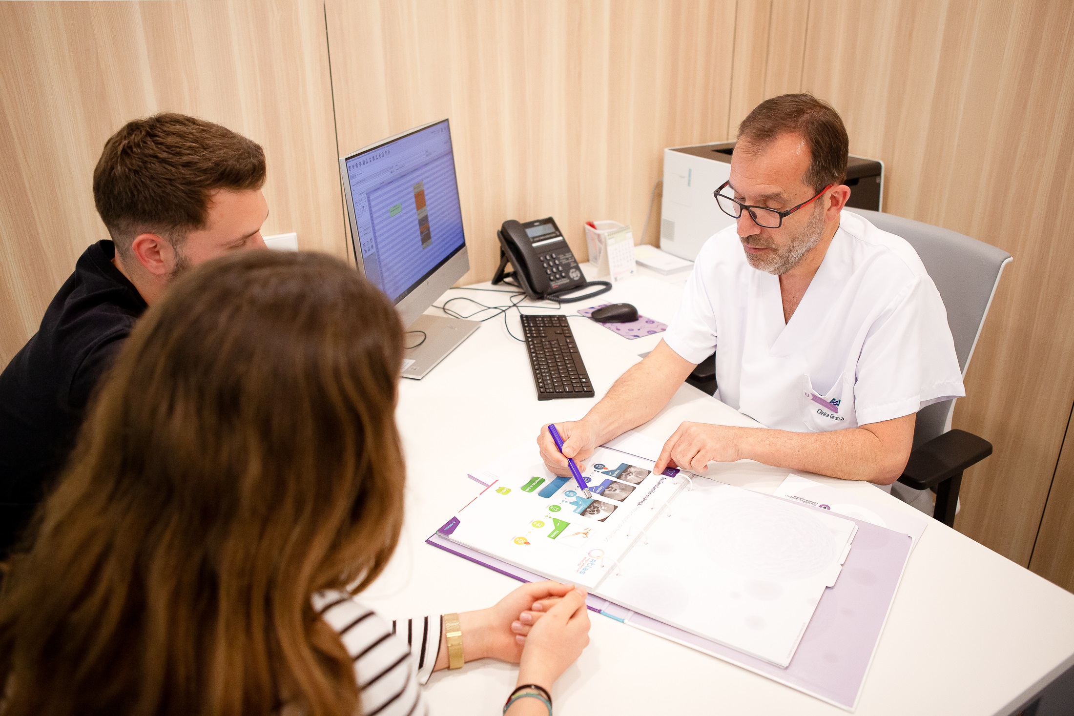
The first step to determine the cause of infertility and apply the optimal treatment is to conduct a couple’s evaluation and review provided medical reports to gather all necessary case-specific information.
However, no single test can conclusively assess a couple’s reproductive potential. Only the woman’s age is a proven prognostic factor, as advanced age reduces the likelihood of natural conception.
Each case is unique, but typically, during a first visit to GIROFIV, the gynecologist will request a basic fertility workup. If more specialized tests are needed, a complementary study is usually recommended.
A detailed anamnesis to understand the patient’s medical background, including:
Assesses the anatomy and functionality of the female reproductive system. An early-cycle ultrasound evaluates ovarian reserve by quantifying antral follicles in each ovary.
Measures key hormones (AMH, FSH, LH, estradiol, progesterone...) to evaluate ovarian function.
Includes evaluation of:
The primary diagnostic tool for male fertility. Evaluates the quality of the sample and macroscopic values (semen volume, appearance, pH, etc.) and microscopic values (concentration, mobility, vitality and morphology).
The study of the number and structure of chromosomes within cell nuclei. Conducted via blood test.
Each body cell should contain 46 chromosomes (22 autosomal pairs + 1 sex chromosome pair). Gametes (eggs/sperm) carry only 23 chromosomes to ensure the embryo’s correct total.
Karyotype abnormalities increase the risk of genetically abnormal gametes, leading to infertility or recurrent miscarriage.
A minimally invasive imaging test using vaginal ultrasound and contrast gel (ExEm Foam) to assess uterine cavity and fallopian tube patency. Less painful than traditional hysterosalpingography and performed in-office. Critical for diagnosing tubal obstruction (30% of infertility cases).
A surgical procedure under general anesthesia using abdominal optics to inspect pelvic organs (uterus, ovaries, fallopian tubes). Detects adhesions or blockages and may include therapeutic interventions.
Direct visualization of the uterine cavity via cervical insertion of an optic device. Diagnoses polyps, fibroids, adhesions, or other endometrial abnormalities.
A molecular test to identify the optimal embryo transfer window by analyzing endometrial gene expression. Requires an endometrial biopsy (no sedation). Indicated for implantation failure with high-quality embryos.
Chromosomal analysis via blood test to detect abnormalities that may cause infertility or recurrent pregnancy loss.
Evaluates DNA integrity in sperm. High fragmentation rates correlate with poor embryo quality and miscarriage risk. Uses CometFertility technology to differentiate single-strand (reduced fertilization) and double-strand (miscarriage risk) breaks.
Causes: Primary (testicular dysfunction) or secondary (varicocele, toxins, chemotherapy, smoking, obesity, age >45).
Analyzes sperm for chromosomal abnormalities (X, Y, 13, 18, 21). Limited to patients with more than 1 million sperm/mL.
Referral to an andrologist for sexual dysfunction, testicular pain, or suspected varicocele.